- Browse Repository
- University Libraries
- Kelvin Smith Library: Projects & Publications
- Art of STEM
- Art of STEM 2016
Art of STEM 2016
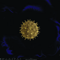
Show morePlants release pollen into the air as part of their reproductive process. Humans breathe in the pollen and the body mistakes it for invading germs, triggering the body's defense, resulting in a sneeze to expel the intruder out. Image was taken using a Helios NanoLab 650 electron microscope at the Swagelok Center for Surface Analysis of Materials. It measures approximately 100 microns in diameter. The single grain of pollen was cut from the full microscope image, colorized using Photoshop and imposed on to a stylized background.
Show less
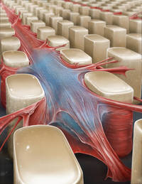
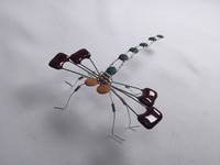
Show moreThe image illustrates a microfabricated three dimensional (3D) substrate to confine, align, and elongate biological cells. Pillar top surfaces are rendered non-adhesive to cells and sidewalls of the micropillars were functionalized to allow adhesion of cells in the inter-pillar space. Biological cells, such as stem cells and heart muscle cells, can be confined in a 3D micropillar environment with precise control over mechanical properties, so cell behavior in response to various mechanical effects can be characterized. I would like to acknowledge my team member Grace Gongaware from Cleveland Institute of Art and my supervisor Dr. Umut Gurkan for contemplating and crafting this image.
Show less
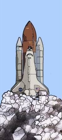
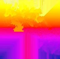
Show moreThis is a postcard for a lecture and film screening I organized on campus last fall. The lecture was by NASA engineer Matt Melis, who spoke on the Columbia accident investigation; the film, screened by the CWRU Film Society, was "Ascent: Commemorating Shuttle," which he produced. (It can be found on YouTube; highly recommended for space and/or photography enthusiasts.) My initial idea for the postcard was in art nouveau style, with an ornate frame featuring text about the event and mission badges. Ultimately I wound up scrapping that, because I had trouble capturing the way it had looked in my head, and because the text began to seem like too much of a distraction. I kept the flat colors and heavy outlines, which made it look more like a stained glass window. This struck me as appropriate, because launch vehicles always exist on a mythical scale. As the shuttle program fades into history, it joins that pantheon, loses its immediacy, and becomes more abstracted in our collective understanding, like a historical figure turned religious icon. I also wanted to show the shuttle frozen in time in a kind of repose, nestled into the clouds - a real contrast with the violence of a launch, but the kind of visual that suggests itself when you watch from a distance - which the edgeless panel is well-suited for. I want the viewer across the room to look at it on a bulletin board (long after the event it advertised is over) and feel themselves press "pause." I hope it worked on you. ------------------------------------------------ Size: 4"x9"
Show less
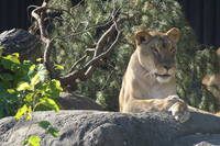
Show moreShe was sitting in the best position for a portrait photo although she kept moving her head. I wanted to get much close, but knew that I was unable. I learned that when taking photos of animals you must take more than one picture because they move when you don't want them too. This was beautiful!
Show less
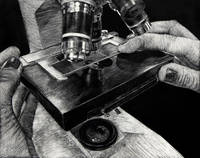
Show moreThis clayboard artwork features an image of my sophomore year biology lab partner's hands working a microscope. Lab days were my favorite days because we were able to use the microscopes. It always fascinated me that humans were able to create such a tool that can look into the cells of plants or analyze the blood stream of fish. By using a clayboard to create my work, I was able to analyze the microscope which in turn was analyzing a slide. Honors Biology with Mrs. Horvath was the first very challenging class I took in high school. She pushed us to work hard and drilled into us the difference between learning and memorization. Mrs. Horvath managed to draw us into the interesting side of biology. "Bio with Horvath" was created to recall those times and remind me and my classmates of those passionate lectures about ATP.
Show less
Show moreHuman motor function is dependent on neuromuscular junction that links nerve system and muscular tissues. The integrity of neuromuscular junction is impaired in many human diseases including amyotrophic lateral sclerosis (ALS), which is well known as the ALS Ice Bucket Challenge. The pathogenesis of ALS is not well understood and there is no cure for this fatal disease. Presented above is a confocal image revealing the structure of neuromuscular junction of transversus abdominis muscle isolated from ALS transgenic mouse. Tissue section was stained using mouse anti-neurofilaments antibody (green) to show nerve fibers in muscle. Nicotinic acetylcholine receptors, which are presynaptic marker to transmit nervous signal to muscular tissues, were stained by dye conjugated bungarotoxin (red). The specimen was imaged with a 40x air objective using spinning disk confocal. Author's seal is presented in Chinese in lower right.
Show less
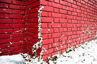
Show moreThis photo was taken on a snowing day the past winter. The color of this Boston-ivy drew my attention when I was walking back to the North Residential Village. Some branches of it were already dead and turned to yellow. The only branch that is still alive was covered with snow, but it still shows its energy to keep growing higher and conquering the bad weather. Boston-ivy seems to die every winter, but it will always come back to live the next spring. Cleveland is cold, but the spirit of Boston-ivy warms our world up.
Show less
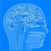
Show moreThis is a digitally drawn cut-away view of a human head with a brain composed of a schematic of several circuits including many resistors, transistors, capacitors, and diodes. This was inspired from the fact that our brains work based on electrochemistry, and electricity flows through the brain just as electricity flows through a circuit. The programs used were Adobe Photoshop, Krita, and Microsoft Paint.
Show less
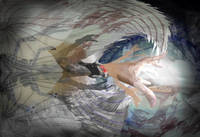
Show moreIncorporation of STEM: By merely using technology, a form of science, I utilized science as a device to create this piece. Primarily computer science was used to create it. I used the program Adobe Photoshop to create this composition. To create the composition, I took myriad photographs and scans to be utilized as frames of the piece, a clear use of technology in the work. The use of technology is also emphasized in the claw that is on the far left side of the composition. I used LEGO pieces to construct the apparatus and extended it to achieve the desired shapes. In turn, a considerable amount of engineering was utilized to make and extend the claw. Mathematics was used to create the transparent architectural sketch on the right side of the composition. Extra Information: This personal map is a culmination of my thoughts of my past and how the future (forcefully) brings them together. The left side of the piece is clearly dimmer, implicating the past. The faded flower at the extreme left is the harbinger of the start of my tolerance for my mixed heritages. The upper tier of the typhoon depicts the first family reunion of my father's lineage: the African American Davis' are shrouded in savanna-like thickets, representing my lack of familiarity with my fraternal heritage. The lower tier of the typhoon depicts a more recent family reunion: me amongst my less alienating Japanese cousins and grandmother. My attachment to this lineage is represented by a fragment of my Japanese language notes. The two tiers are divided by my radiated arm emerging from claws which propel it to the present and future. Additional appendages sprout in protest, depicting my inner struggle to tolerate a change of my cultural idiosyncrasy. Into my unknown future, a light of change is not grasped by the arm, but it instead absorbs the arm and the typhoon into one unstable yet defined focus. Amid the turbulence, the two landscapes of the left switch places and witness the blend. They fade into the background, omnipresent yet unobtrusive in the light of my future career of architecture. One of my conceptual sketches blends in with the harmonious families, implying that my newfound tolerance will undoubtedly benefit my success in later years.
Show less
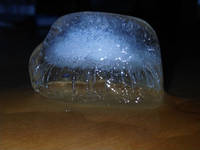
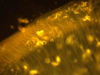
Show moreThe variations in crystal structure within this melting ice diffract light in different ways, such that an opaque cloud appears to be trapped inside this melting ice cube. This "cloud," a formation normally suspended in the air, is here suspended instead in a solid block of ice.
Show less
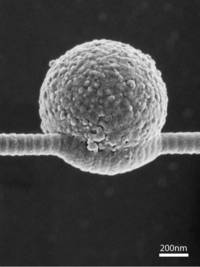
Show moreDepicted are individual collagen fibers extracted from the tissue of the tail of the rats in solution. Collagen is a protein found in the connective tissue between cells and is one of the most abundant proteins in the human body, with estimates of 25 to 35 percent of all protein content. This picture was taken by means of dark field microscopy, a special technique that must be used due to the fact the collagen fibers would be impossible to see in solution using ordinary bright field microscopy. This image was taken with help from Yehe Liu and was part of an effort to better understand the micro level mechanical characteristics of an individual collagen fibril.
Show less
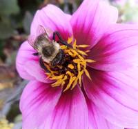
Show moreI wasn't just taking random pictures, I actually used my resources. It was fairly easy and a lot of fun trying to dodge the bees. It took about five tries to get the one I actually wanted. I had to position the camera vertically, bend down and move in close, to capture the angle that I wanted. The only thing that was difficult for me were the bees flying around me. It made it very uncomfortable to capture the picture I wanted, yet easy, too.
Show less
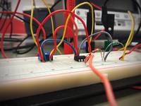
Show moreJust a simple iPhone 6 photo of our ENGR 210 Circuits Lab 5 assignment. This picture is the result of my team's hard work and hours of stress, struggle, laughs, more stress, more struggle, more laughs and final success. Thank you, Jenny and Helen, for being awesome. Modified with Snapseed App for iPhone.
Show less
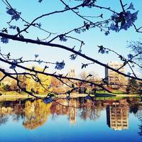
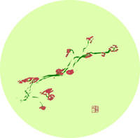
Show moreWhen I walked along the lake and spotted this beauty of nature reflected on the lake, I was mesmerized by how beautiful our nature was. As an engineering major, I appreciated science (biology in this case) for creating all different colors. Also, the reflection on the lake even made me think of my own life.
Show less
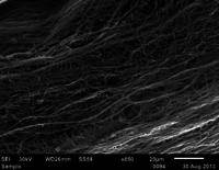
Show moreThe image considers unique polymeric fibers that fabricate through a novel melt co-extrusion process developed at CWRU. Most fiber production methods make random fibers resembling cooked spaghetti. Our interesting fiber-making process will develop new aspects for fiber applications in many areas. This work was a collaboration with E. Baer and G. E. Wnek.
Show less
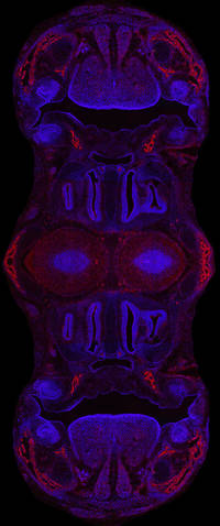
Show moreThis image was compiled from many photos taken on a digital microscope camera that uses ultraviolet light to bring out blue and red fluorescent signals. The compiled images were then mirrored and merged for design purposes in Photoshop CC. Blue marks the nuclei of cells and red marks bone precursor cells. This modified image comes from a section of normal embryonic mouse skull, which showcases the nasal cavity, mouth, teeth, and tongue. The Atit lab typically uses sections like these from normal mice to comparatively identify abnormal skull structures (morphology) in genetically modified mice. These images are then used to study the development of cranial bone and cartilage in mice as a model for human development.
Show less
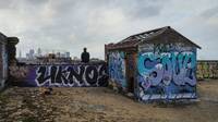
Show morePerched atop a piece of Cleveland's past, it's hard not to think of its future. This photograph was taken on the roof of the Warner and Swasey Company building. This company was, in its day, well known for its production of astronomical telescopes. The telescope that is currently on the roof of the A.W. Smith Building at Case Western Reserve University was built by them in 1894. Warner and Swasey may have left the city decades ago, but the building remains as a testament to Cleveland's industrial and scientific past as the city moves to create a new future. This photograph was taken by Neal Mathes in December of 2015 while urban exploring with a friend.
Show less