- "Steele, Miarasa" (x)
- Search results
Search results
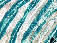
Show moreThis photograph was taken with a digital microscope camera and the colors modified for aesthetic appeal using Photoshop CC. The image shows overgrown hair follicles and surrounding skin cells in a tissue sample from a mouse with skin fibrosis. The deep blue cells constitute the epithelial lining of the hair follicle shafts, and the pale tissues are the hair shafts proper. This image represents several years of work that the Atit Lab has put into successfully developing a transgenic mouse model for finding therapeutic treatments for skin fibrosis. This image won first place in the CWRU category.
Show less
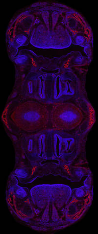
Show moreThis image was compiled from many photos taken on a digital microscope camera that uses ultraviolet light to bring out blue and red fluorescent signals. The compiled images were then mirrored and merged for design purposes in Photoshop CC. Blue marks the nuclei of cells and red marks bone precursor cells. This modified image comes from a section of normal embryonic mouse skull, which showcases the nasal cavity, mouth, teeth, and tongue. The Atit lab typically uses sections like these from normal mice to comparatively identify abnormal skull structures (morphology) in genetically modified mice. These images are then used to study the development of cranial bone and cartilage in mice as a model for human development.
Show less