- Browse Repository
- University Libraries
- Kelvin Smith Library: Projects & Publications
- Art of STEM
- Jennings, Wayne (x)
- Art of STEM 2015
Art of STEM 2015
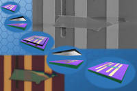
Show more2D materials are of great attraction for their exotic properties. The flow in the image shows a facile way to manipulate these nano-scale materials. The manipulation is assisted by a PDMS stamp with the help of a micro-positioner. The optical microscopic (lower-left) and SEM (upper-right) images of a fabricated 2D material device are shown. This manipulating technique helps scientists and engineers explore the characteristics and applications of these promising materials.
Show less
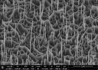
Show moreVertical ZnO 3D nanostructures were synthesized on Au covered c-plane GaN epilayer film on sapphire substrate by chemical vapor deposition method. The growth was conducted in a horizontal tube furnace with 1" diameter quartz tube. High purity ZnO powder and oxygen were used as the precursors. The growth was carried out for 1 hr at 900 C. The growth direction was polar. The image shows the 20 degree tilted view of the nanostructures. The image was taken at the Swagelok Center for Surface Material Analysis (SCSAM) at CWRU.
Show less
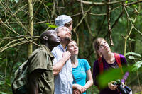
Show moreDr. Rollins, Daniel Hageman, Alexander Kolberg and Nicole Mammoser are observing chimpanzees on a brief excursion to Uganda in order to assess medical needs.
Show less
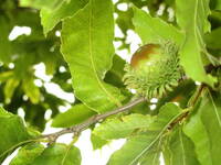
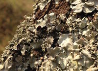
Show moreI was walking along the path around the duck pond in front of the Cleveland Museum of Art, looking to capture life along with other plants and flowers. I came upon this interesting object hanging from the tree. The flash really helped to lighten up the entire composition.
Show less
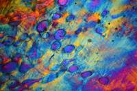
Show morePolarized light microscopy of mechanically aligned, electrochemically compacted collagen. Collagen molecules are isolated from skin samples and processed into a semi-viscous fluid. This fluid is then introduced into a mold between two carbon electrodes. By applying a voltage across the electrodes, a pH gradient is formed. The collagen molecules are driven into a thin sheet or thread, which can then be used as a scaffold for cell culture studies and development of engineered tissue. These scaffolds can be stretched in order to align the molecules, which in turn will induce any cells seeded on the scaffold to align. By varying the amount of stretching, varying degrees of alignment can be achieved. In the polarized light microscopy image "Order in Organic Chaos" a partial alignment of the scaffold is presented. The blue areas indicate alignment along the diagonal of the image running from the bottom left to the top right, whereas the other shades represent localized misalignment. The elliptical shapes are the result of gas bubbles, released during electrical compaction, being trapped in the sheet. By varying the time of compaction the presence of such bubbles can be limited. And so, from a vial of randomly floating molecules, ordered structures can be formed and controlled.
Show less
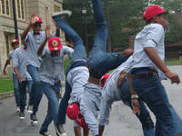
Show moreThis image was created as we studied how to capture action in our photography. We experimented with a variety of activities and actions, and captured a lot of energy in motion. We had to break down that motion and capture it on film. The final image had to show the energy in one photograph and, with the help of Photoshop, this image presented itself. Print size: 8x10
Show less
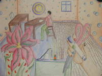
Show more"My Contour Nouveau piece is an overall drawing about what we've been doing in class. In my piece you can see a kid sitting at a table working on his own artwork and another kid's hand holding up things so she can draw her own hand. I selected five different cellular designs to incorporate into my piece. They include red blood cells, stem cells, pansy petal cells, wood cells and streptococcus cells. I chose the red blood cells to pattern the flower pot because the flower pot was green. Green and red are complimentary colors and look good together. I chose the stem cells because they looked different and made the walls stand out. I chose the pansy petal cells because I wanted the flower petal to be made of flower cells. I chose the wood cells for the floor, as it made sense. Finally, I chose the streptococcus cells to design the boy's pants because I had gotten strep the week before we started our Art Nouveau project and I wanted to know what the virus looked like."
Show less
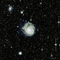
Show more"A digital photograph of the Galaxy Messier 101 (also known as the Pinwheel Galaxy), constructed over the course of three (non-consecutive) years using the Burrell Schmidt Telescope at Kitt Peak National Observatory. The galaxy was imaged four times, using four different color light filters: a filter that isolates mostly blue light, one that isolates greenish light, one that isolates mostly red light, and one that isolates a very specific kind of red light emitted mostly by newborn stars. Each of the four images contains around 20 hours of total exposure time. This color image of the galaxy was constructed by layering five different images: the red, green, and blue filter images to create an approximately true-color picture of the galaxy and surrounding environment, an image in which the newborn stars were isolated from the background starlight (seen as the pockets of red throughout the galaxy's spiral arms and elsewhere), and an image of the galaxy that has been heavily processed in order to bring out the faintest starlight at the galaxy's edges (seen as the faint blue 'halo' of light surrounding the galaxy). The latter contains stars that were most likely torn away from the Pinwheel through gravitational interactions with one or more of its neighbors (possibly the smaller galaxy seen near the bottom of the image), and even some very young stars that may have formed through the collapse of extended gas and dust (not pictured) caused by the interaction. The faintest light seen in this image is 1000 times fainter than the dark night sky. Observations of M101 were done by the following people: Chris Mihos, Paul Harding, Craig Rudick, John Feldmeier, Chelsea Spengler, and Aaron Watkins."
Show less
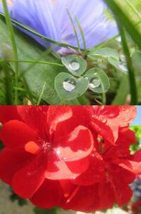
Show moreAs I looked closer at taking images of plants and flowers, I had to make sure I was choosing the right angle for taking photos of flowers, which is the biggest issue because there are so many. To be honest, I took the photo of whatever caught my eye. I wanted the picture to grasp emotion as much as possible. As I worked with other students, I found my ideas were different from others' ideas and they wanted to replicate an image they saw while I just made my own ideas. My epiphany was, if you get the right angle with the sun and with the water droplets you could get a sparkle on the flower petals. I learned when taking plant and flower photos, choosing the right angle is very difficult.
Show less
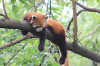
Show more"Noon on a Sunday afternoon, an Ailurus fulgens (Red Panda) wakes from a day nap astride Morus nigra (Blackberry) tree some 15 meters high. Rest is both relaxing, hunting, and camouflage. The subtle fur changes color along the ventral with the dorsal planes exhibiting a delightful contrast. The top fur resembles the red and dark brown soil; the lower coat blends with the darker tones of bark and shaded earth. With keen senses of smell, hearing, and eyesight Ailurus fulgens can identify components of its omnivorous diet atop the branches. Ultimately however, the red panda's draped limbs counter balance with its tail allowing carefree napping. 6/15/2014, Chengdu Research Base of Giant Panda Breeding, Chengdu, Sichuan, People's Republic of China"
Show less
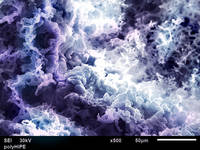
Show moreHigh internal phase emulsions (HIPE) are materials with incredible microscale morphologies. They normally exhibit a pore like structure with polymerized scaffolding surrounding spherical voids. This portion of the sample was somewhat different however, it illustrates how the structure changes under deformation and how the polymer reacts to non-reversible strain. This image was taken using a scanning electron microscope (SEM) with a sample sputter coated in a fine layer of gold. Gold makes the sample conductive so that it may scatter incoming electrons from a concentrated electron beam to a detector thus creating an image.
Show less
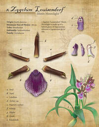
Show moreA botanical plate illustration highlighting the detail of the orchid xZygolum Louisendorf from South America. Created using Adobe Photoshop and Illustrator.
Show less
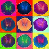
Show moreThis is one transverse section of a wild type, or genetically "normal," rodent spinal cord I imaged at 16x magnification and presented in nine screens, à la Warhol. The spinal cord section was originally stained using a technique called 'Immunohistochemistry' to look for a subpopulation of neurons that express the neurotransmitter serotonin. I then used Photoshop to artificially color the sections and to hand draw the supporting cells as well as the many possible neural connections it's able to make. The intent of this piece is to show how incredibly important the use of colors are to neuroscientists. We use colors to identify and highlight important players involved in Central Nervous System physiology with the expressed purpose of finding out where these important players are, when and how they interact with themselves as well as other neurological elements in vivo. Using an array of colors bound to specific proteins, we can therefore begin to elucidate certain aspects of how our Central Nervous System works.
Show less
Show morePicture captured of a praying mantis I captured in Rwanda, Africa. By far the most well-camouflaged organism I have ever encountered. Be sure to look carefully for the insect. Usually people first see the leg, which you can follow up to the rest of the mantis. There are very few representatives of the genus Oxylea in collections anywhere in the world, because no one can find them. This is probably a new species, and will be described and published in the next year or two.
Show less

Show moreThe intended artwork is not the submitted compiled photographs, but it is the subject of the photographs instead. The sculpture is a laptop computer made out of LEGO pieces, approximately 1,000 - 2,000 pieces. Its dimensions are a 27 centimeter width, 37.7 centimeter length, and 3.9 centimeter height. In the photograph of the top left corner, the artwork (right) is compared to its model, a Windows 7 home use laptop. I managed to create the artwork by using the model and observing some of its key features. Originally, the artwork was meant to be a replica of the model, but a lack of maroon LEGO pieces allowed for creative liberties. The artwork's labeled keyboard (bottom left and bottom right photographs) lies just beneath the artwork's main attraction: the screensaver. The "opened" artwork (top center photograph) shows off its screensaver: the logo of the laptop's Internet browser, Google Chrome, a variety of desktop applications, and the phrase "STEM Builds." The closed artwork has a generally white design with purple stripes and a couple of maroon stripes, reminiscent of its model's cover color. Just as I was able to build a laptop out of LEGO pieces, STEM allows people to create anything from buildings to home appliances. So, in tune with this fact, the artwork represents anyone's desire to create. I managed to build a laptop with STEM, so what will STEM help you build?
Show less
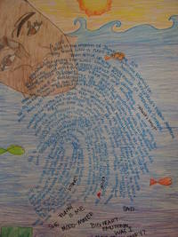
Show moreI created this image after studying about the Forensic Sciences. One of the areas of Forensics that we learned was about fingerprinting; what they look like and what they mean to us personally. This fingerprint is mine along with the story that goes with it. That's why it is personal to me and no one else. This entry won an honorable mention for the high school category.
Show less
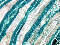
Show moreThis photograph was taken with a digital microscope camera and the colors modified for aesthetic appeal using Photoshop CC. The image shows overgrown hair follicles and surrounding skin cells in a tissue sample from a mouse with skin fibrosis. The deep blue cells constitute the epithelial lining of the hair follicle shafts, and the pale tissues are the hair shafts proper. This image represents several years of work that the Atit Lab has put into successfully developing a transgenic mouse model for finding therapeutic treatments for skin fibrosis. This image won first place in the CWRU category.
Show less