- Browse Repository
- University Libraries
- Kelvin Smith Library: Projects & Publications
- Art of STEM
- Art of STEM 2015
Art of STEM 2015

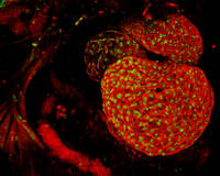
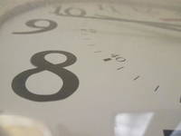
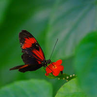
Show moreThis image was created as we studied how to capture action in our photography. We experimented with a variety of activities and actions, and captured a lot of energy in motion. We had to break down that motion and capture it on film. The final image had to show the energy in one photograph and, with the help of Photoshop, this image presented itself. Print size: 8x10
Show less
Show moreThis picture illustrates neurons regenerating though a spinal cord injury via a bridge made of peripheral nerves. To facilitate axonal regeneration, many treatments must be combined to create the "Full Monty." The inhibitory glial scar is broken down with an enzyme, a bridge is built using peripheral nerves, and the integration of the bridge and spinal cord is encouraged with growth factors. Axons in yellow can be seen leaving the blue spinal cord, regenerating through the bridge, and reentering the spinal cord. This image uses antibodies against tyrosine hydroxylase and glial fibrillary acidic protein imaged at 20x via confocal microscopy.
Show less

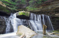
Show moreThe Great Falls of Tinkers Creek is a waterfall located in Viaduct Park, Bedford, Ohio. These impressive 15-feet falls flow through an old mill and viaduct.
Show less
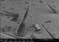
Show more"SEM image of a NiTi fractured surface that resulted from flex bending fatigue, 800x magnification (center image). Image is saved as 30x30 inches but can be reduced as necessary."
Show less
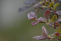
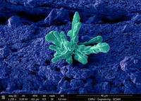
Show moreThis photo shows a rather waxy plant whose surface caused water droplets to remain separated on its surface. The photo was taken using a Nikon D7100 with 300m lense.
Show less
Show moreIndium nitride (InN) nanowires were synthesized on Si (100) substrate by chemical vapor deposition (CVD) method via a vapor-liquid-solid growth mechanism. The Si substrate was covered with 15 A Au film deposited via thermal evaporation. High purity Indium foil and ammonia were used as group III and group V sources respectively. The growth temperature and pressure were 580 C and 10 Torr respectively. The growth was conducted for 1.5 hr. The SEM image shows two different orientations of the InN nanowires; non polar m-plane with triangular cross section and polar c-plane with hexagonal cross section. The image was taken at the Swagelok Center for Surface Analysis of Materials (SCSAM) at CWRU.
Show less
Show moreWe were studying how to manipulate the aperture on our camera and to make the depth of field short and create an image that has dramatic blur. I chose this as my best image because it took me a long time to finally get a decent picture and learn how to take the photo right. Print size: 8x10
Show less

Show moreBeing able to manipulate your shutter speed makes picture taking easier because, if you need a clearer photo you can speed up the shutter speed. If you want a blur you can slow it down. I needed a fast shutter speed to be able to capture Keyshauna in action. She looks as if she is just hanging there, in flight.
Show less
Show moreThis is a Silicon Carbide crystal growing on top of Silica glass. The image was taken using an FEI Helios 650 high resolution scanning electron microscope at the Swagelok Center for Surface Analysis of Materials. The image was acquired using secondary electrons at relatively low voltage (5kV) to boost contrast and a tilt angle of 52 degrees to enhance the 3D feel of the surface.
Show less
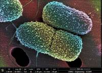
Show moreSEM autograph of Pseudomonas aeruginosa: a ubiquitous, gram-negative, rod-shaped bacterium. It has been intensively studied as an opportunistic human pathogen. It is one of the most common pathogens for nosocomial infection in immunocompromised individuals. This image was taken with FEI Helios Nanolab 650 using 2 KV 50 pA with TLD mode 2. The specimen was prepared with the OTO technique.
Show less
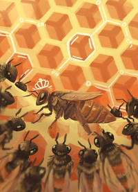
Show moreThis piece is intended to show the effects of the queen and the mandibular pheromone she emits to the worker bees and to highlight the parallels between the ring structure in methylparaben, a key component of Queen Mandibular Hormone, and the hexagonal honeycombs that make up the bee hive. The piece is 13 x 18 inches
Show less
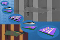
Show more2D materials are of great attraction for their exotic properties. The flow in the image shows a facile way to manipulate these nano-scale materials. The manipulation is assisted by a PDMS stamp with the help of a micro-positioner. The optical microscopic (lower-left) and SEM (upper-right) images of a fabricated 2D material device are shown. This manipulating technique helps scientists and engineers explore the characteristics and applications of these promising materials.
Show less

Show more"The Marsupial Mother: Embryonic Diapause in Macropus rufus" is intended to educate viewers of the female kangaroo's reproductive adaptations. The illustration depicts the anatomy of the reproductive organs, as well as the development of an embryo into a joey. By clearly showing the life stages of the newborn, this piece visually conveys a large quantity of information that is readable for the viewers. The project design focuses on a specimen's adaptation rather than an emphasis on the animal in general. This piece effectively uses the two, filled line art insets, as well as the realistic rendering of the mother and joey kangaroo to communicate how the adaptation pertains to the subject. Through its visual aesthetic and the subject matter, this project is able to inform, intrigue, and stimulate curiosity, ultimately finding a unique approach to education.
Show less
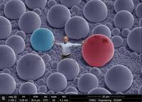
Show more"The picture is a Secondary Electron image on the FEI Helios 650 combined with an iPad photo. The sample in the SE image is a Tin balls specimen used for system alignment. The imaging was done at ""Swagelok Center for Surface Analysis of Materials"" (SCSAM) in the Case School of Engineering."
Show less

Show moreA group of silica microspheres are "dancing" to high-frequency "music" of a multimode, micromechanical plate resonator in an aquatic environment. The magical two dimensional (2D) geometric patterns, namely, Chladni figures, visualize the multiple flexural modes when microspheres are stabilized along nodal circles and nodal lines. This is the first time we resolved such microscale Chladni figures on a silicon carbide trampoline resonator, and such techniques may enable single-cell manipulation, cell patterning, size-based cell sorting and related cellular and biophysical studies.
Show less