- Browse Repository
- University Libraries
- Kelvin Smith Library: Projects & Publications
- Art of STEM
- Art of STEM 2016
Art of STEM 2016

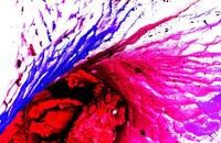
Show moreWhen I walked along the lake and spotted this beauty of nature reflected on the lake, I was mesmerized by how beautiful our nature was. As an engineering major, I appreciated science (biology in this case) for creating all different colors. Also, the reflection on the lake even made me think of my own life.
Show less
Show moreHuman motor function is dependent on neuromuscular junction that links nerve system and muscular tissues. The integrity of neuromuscular junction is impaired in many human diseases including amyotrophic lateral sclerosis (ALS), which is well known as the ALS Ice Bucket Challenge. The pathogenesis of ALS is not well understood and there is no cure for this fatal disease. Presented above is a confocal image revealing the structure of neuromuscular junction of transversus abdominis muscle isolated from ALS transgenic mouse. Tissue section was stained using mouse anti-neurofilaments antibody (green) to show nerve fibers in muscle. Nicotinic acetylcholine receptors, which are presynaptic marker to transmit nervous signal to muscular tissues, were stained by dye conjugated bungarotoxin (red). The specimen was imaged with a 40x air objective using spinning disk confocal. Author's seal is presented in Chinese in lower right.
Show less
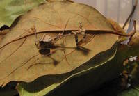
Show morePraying mantis nymph (Sibylla pretiosa) demonstrating the cryptic effect achieved by cuticular expansions of the exoskeleton. This species lies in wait on trees and leaves for unsuspecting prey. We found this specimen imitating a leaf blowing in the wind by waving her arms and abdomen in a single direction. Taken in Rwanda, Africa 2014.
Show less
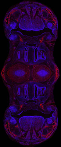
Show moreThis image was compiled from many photos taken on a digital microscope camera that uses ultraviolet light to bring out blue and red fluorescent signals. The compiled images were then mirrored and merged for design purposes in Photoshop CC. Blue marks the nuclei of cells and red marks bone precursor cells. This modified image comes from a section of normal embryonic mouse skull, which showcases the nasal cavity, mouth, teeth, and tongue. The Atit lab typically uses sections like these from normal mice to comparatively identify abnormal skull structures (morphology) in genetically modified mice. These images are then used to study the development of cranial bone and cartilage in mice as a model for human development.
Show less
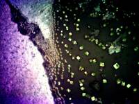
Show moreThis image captures the transition in time of a perovskite reaction, a material used in solar devices. The intersection of reacted perovskite, left, and unreacted tin iodide crystals in a matrix of methylammonium solution (MAI), right, highlight the process of creating perovskite, as the unreacted region on the right shows large crystals on the order of several microns decreasing in size as the crystals get closer to the reacted perovskite region on the right. Tin iodide crystals appear to burst out of the perovskite that it was made from, when in fact the two regions were spin coated 20 seconds apart, and thus could not react. From this image, we elucidate that the reaction time for perovskite formation is incredibly fast, and we are closer to understanding the mechanisms for perovskite reaction.
Show less
Show moreFollowing excitation by light, molecules try to find the quickest and easiest path to relax. However, sometimes this leads to trapping in a minimum rather than getting back to where it started.
Show less
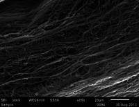
Show moreThe image considers unique polymeric fibers that fabricate through a novel melt co-extrusion process developed at CWRU. Most fiber production methods make random fibers resembling cooked spaghetti. Our interesting fiber-making process will develop new aspects for fiber applications in many areas. This work was a collaboration with E. Baer and G. E. Wnek.
Show less

Show morePerched atop a piece of Cleveland's past, it's hard not to think of its future. This photograph was taken on the roof of the Warner and Swasey Company building. This company was, in its day, well known for its production of astronomical telescopes. The telescope that is currently on the roof of the A.W. Smith Building at Case Western Reserve University was built by them in 1894. Warner and Swasey may have left the city decades ago, but the building remains as a testament to Cleveland's industrial and scientific past as the city moves to create a new future. This photograph was taken by Neal Mathes in December of 2015 while urban exploring with a friend.
Show less
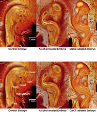
Show moreQuail embryos at day 3 of development (similar to human week 5). Left is a normal control embryo that has a straight trunk and the head curves nicely to the right. Middle is an embryo treated with alcohol at an earlier time point (day 1, similar to human week 3). This is mimicking fetal alcohol syndrome in human. Right is an embryo in which the cardiac neural crest cells were ablated. This is mimicking DiGeorge Syndrome. Both abnormal embryos show twist and/or rotation of the body. (Work also has contribution from Lindsy Peterson, a former lab member. Adviser: Andrew Rollins) I attached an image with two copies of the same sets of images, the bottom ones have labels on them pointing out some structures of the embryo as part of my description to orient the observer during the judging process. The top part is my participating image.
Show less
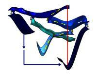
Show moreFickian diffusion, an established law of physics for gas molecules permeating through a material, has been often conceived as a negligible effect for many applications utilizing fluid flow and gas transport. However, with this novel microscale device, Fick's Second Law of Diffusion has been effectively used to control O2 and CO2 concentrations of blood from sickle cell disease (SCD) patients through rapid diffusion. SCD, a notorious and first-found molecular disease, affects the red blood cells' (RBCs) shape and adhesion properties when deoxygenated. This illustration shows the effect of deoxygenation in RBCs as they go through the micro-gas exchanger, sickling and adhering to the specially functionalized microscale channel. This work was a collaboration with Grace Gongaware, a Cleveland Institute of Art student, drawn with Adobe Photoshop and Illustrator, and advised by Dr. Umut Gurkan.
Show less
Show moreA Scanning electron microscope was used to analyze the lead and tin alloy created in Materials Lab (Under Professor Lagerlof). Photoshop was used to add colors to the impurities and grain boundaries to make the alloy seem like a kaleidoscope (K(alloy)scope).
Show less
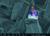
Show moreImages of Gallium Oxide (courtesy of Hongping Zhao and Subrina Rafique) remind me of the fortress of solitude of that famous superhero from Cleveland/Krypyon.
Show less
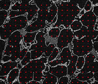
Show moreAn image taken by a scanning electron microscope of a lead-tin alloy, similar to old-fashioned plumber's solder. The white regions correlate to lead while the black regions are tin. Contrast has been augmented, highlighting the complex concert of metals. The main phase sections have been proportionally measured using a grid red dots. This will be helpful for finding the recipe used to create the alloy.
Show less
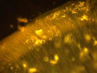
Show moreDepicted are individual collagen fibers extracted from the tissue of the tail of the rats in solution. Collagen is a protein found in the connective tissue between cells and is one of the most abundant proteins in the human body, with estimates of 25 to 35 percent of all protein content. This picture was taken by means of dark field microscopy, a special technique that must be used due to the fact the collagen fibers would be impossible to see in solution using ordinary bright field microscopy. This image was taken with help from Yehe Liu and was part of an effort to better understand the micro level mechanical characteristics of an individual collagen fibril.
Show less
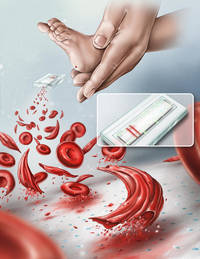
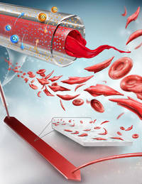
Show moreAbout 3 million people worldwide suffer from sickle cell disease (SCD), mostly in Africa, India and the Middle East, with an estimated 100,000 affected in the US, according to the Centers for Disease Control and Prevention. SCD affects 1 in 375 newborns in the US. More than 800 children are born with SCD every day in Africa, and more than half of them die in childhood due to lack of diagnosis and early treatment. In the US, hemoglobin screening of newborns is mandated for early diagnosis, so that, monitoring and treatment can be started immediately, which has dramatically reduced SCD related mortality. However, this strategy has not been widely available in Africa and other third-world countries, due to limited resources. Therefore, there is a need for simple, rapid, and mobile analyses of hemoglobin types in newborn blood with which to diagnose hemoglobinopathies while the baby is still on-site. Early diagnosis through new born screening in resource-poor settings can be achieved with a point-of-care screening method, such as with the HemeChip, a device developed in the CASE Biomanufacturing and Microfabrication Lab. A HemeChip that can accurately identify hemoglobin type in a drop of blood will improve the way we screen, diagnose and initiate management of hemoglobinopathies in newborns. This illustration depicts the usage of the HemeChip and the process of diagnosing SCD. Collaboration with Umut Gurkan, Yunus Alapan, and James Kim.
Show less
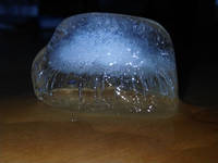
Show moreThe variations in crystal structure within this melting ice diffract light in different ways, such that an opaque cloud appears to be trapped inside this melting ice cube. This "cloud," a formation normally suspended in the air, is here suspended instead in a solid block of ice.
Show less